Radiology Unit

Radiology Center

The A Life Health Group Radiology Unit is equipped with state-of-the-art medical imaging technologies, providing reliable services throughout diagnostic and treatment processes. As a core component of our healthcare services, radiology enables accurate and timely evaluation of our patients’ health conditions. Combining advanced imaging systems with our team of experienced specialists, we are committed to delivering the highest standards of quality in patient care.

Unit Features

Specialist Radiologists

Our team of highly experienced and specialized radiologists meticulously analyzes imaging results to ensure accurate diagnoses and supports healthcare professionals in developing effective treatment plans. Throughout all stages, we maintain a patient-centered approach.

Fast and Reliable Results

The A Life Health Group Radiology Unit ensures the prompt reporting of imaging results, expediting our patients’ treatment processes. Our findings are shared with healthcare professionals in a timely manner, providing the necessary information for immediate interventions when required.

Patient Comfort

We take all necessary measures to ensure the comfort of our patients during radiological procedures. Our user-friendly equipment and experienced staff work diligently to minimize any discomfort throughout the process.

Education and Information

We provide comprehensive information to ensure that our patients are well-informed about radiology procedures. Both before and after the examination, we guide them through each stage, helping them feel comfortable and confident about what to expect.

Advanced Imaging Technologies

Our Radiology Unit is equipped with state-of-the-art devices, including ultrasound, X-ray, magnetic resonance imaging (MRI), and computed tomography (CT). This allows us to provide the highest quality imaging services tailored to the diverse diagnostic and treatment needs of our patients.

MRI – Magnetic Resonance

At Our Ankara Hospital:

The 1.5 Tesla MRI system is among the most widely used and reliable devices in magnetic resonance imaging. This technology leverages a strong magnetic field in combination with radio waves to produce high-resolution images of soft tissues, organs, bones, and other anatomical structures with exceptional clarity.

Unit Features

  1. High-Quality Imaging: The 1.5 Tesla MRI provides detailed, high-resolution images, playing a crucial role in the early detection of various conditions. It delivers clear and sharp visuals that facilitate accurate diagnosis in clinical settings.

  2. Extensive Imaging Options: Our MRI unit offers a wide range of imaging capabilities for evaluating the brain, spine, musculoskeletal system, joints, and abdominal organs.

  3. Patient Comfort: Ensuring patient comfort is a priority. The 1.5 Tesla MRI features a patient-friendly design with a wider imaging space, allowing scans to be performed with minimal feelings of claustrophobia.

  4. Fast and Efficient Results: MRI scans are processed promptly, accelerating treatment planning. Acquired images are rapidly assessed and shared with healthcare professionals for timely intervention.

  5. Expert Radiologists and Technical Team: Our specialized radiologists and experienced technical staff guide patients throughout the imaging process, providing detailed information and ensuring patients feel informed and secure at every step.

At Our North Ankara Hospital:

The 3 Tesla MRI system is one of the most advanced and reliable tools in magnetic resonance imaging. Utilizing a strong magnetic field combined with radio waves, it generates high-resolution images of soft tissues, organs, bones, and other anatomical structures with exceptional precision.

Unit Features

  1. High-Quality Imaging: The 3 Tesla MRI produces highly detailed images, supporting the early detection of a wide range of diseases. Clear and sharp visuals aid accurate diagnoses in clinical practice.

  2. Extensive Imaging Options: This MRI unit accommodates evaluations of the brain, spine, musculoskeletal system, joints, and abdominal organs, providing comprehensive diagnostic capabilities.

  3. Patient Comfort: Designed with patient well-being in mind, the 3 Tesla MRI offers a spacious imaging environment, allowing patients to undergo scans without experiencing claustrophobia.

  4. Fast and Efficient Results: MRI scans are completed quickly, facilitating faster treatment decisions. Images are promptly evaluated and shared with medical professionals.

  5. Expert Radiologists and Technical Team: Our team of skilled radiologists and technical specialists ensures a seamless imaging experience, providing detailed guidance and fostering a sense of safety and confidence for all patients.

1722288399393_634f5a5b_8e408c4b.jpg

CT-Scan

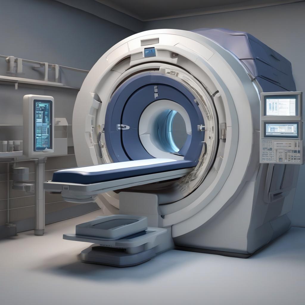
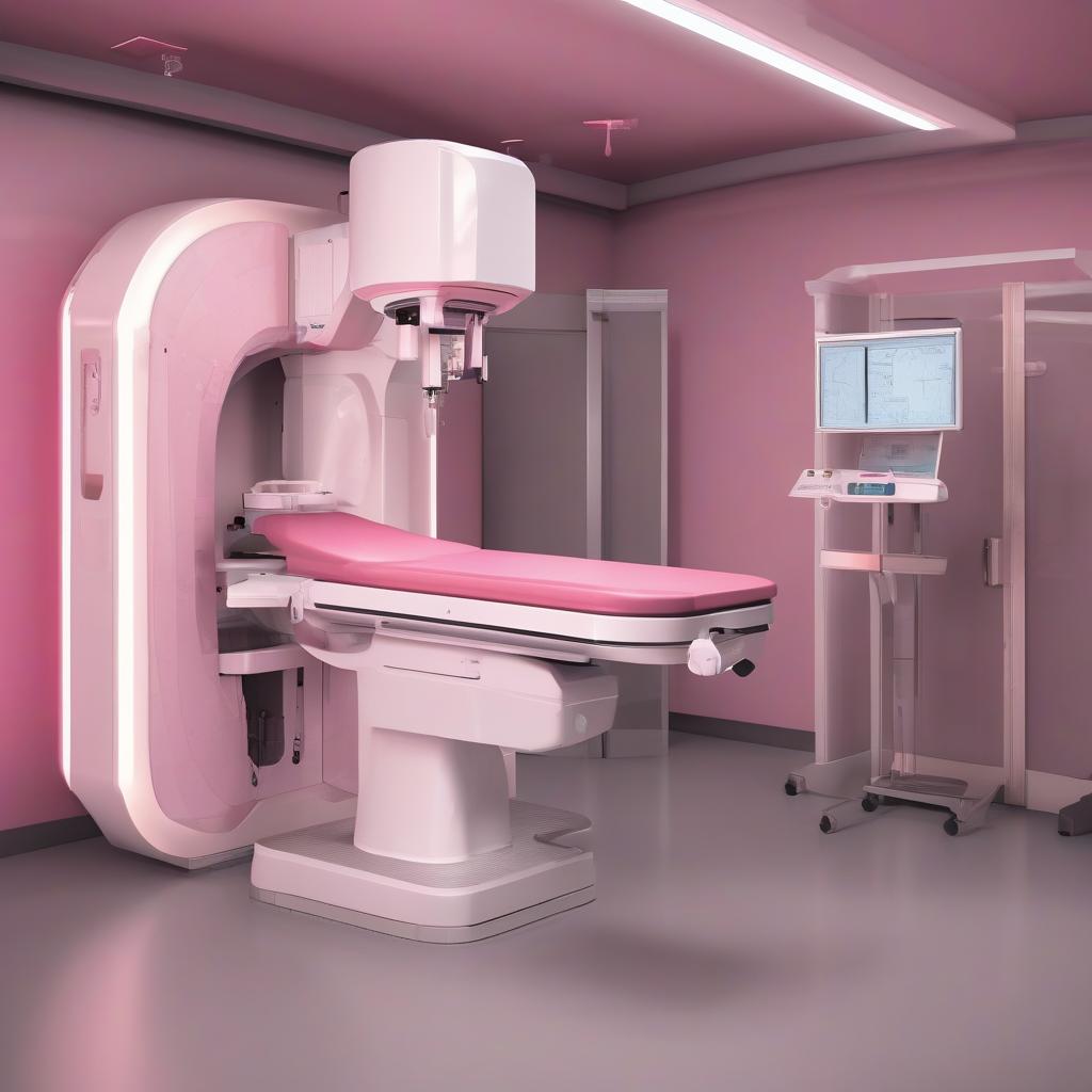
At A Life Health Group Ankara Hospital;

Adopting the aim of providing modern healthcare services, it offers the best service to its patients with facilities equipped with the latest technological medical devices. In this context, our 128-slice computed tomography (CT) device brings together the most advanced technologies to accelerate diagnostic processes and obtain accurate results.

What is 128-Slice CT?

128-slice CT is an imaging technique that allows rapid and high-resolution acquisition of three-dimensional images of the body’s internal structures by taking slices from multiple angles. This technology provides reliable and detailed information for examining all body systems, helping doctors make accurate diagnoses.

Unit Features

  1. High Speed and Image Quality: Our 128-slice CT device performs scans at high speed, obtaining clear and detailed images in a short time. This allows immediate intervention in emergency situations.
  2. Wide Imaging Area: The device enables imaging of many regions such as the head, chest, abdomen, and pelvis, making it possible to examine a wide range of pathological conditions.
  3. Low Radiation Dose: Thanks to its advanced technology, the 128-slice CT provides patients with lower radiation exposure, offering a safe examination process.
  4. Easy and Comfortable Procedure: The scanning process is carried out quickly and comfortably for our patients. Throughout the process, our expert technicians guide and support patients when necessary.
  5. Expert Evaluation: The obtained images are carefully examined by experienced radiologists, contributing to accurate diagnosis and treatment planning.

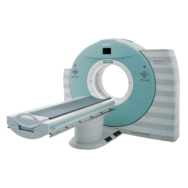
At A Life Health Group North Ankara Hospital;

Adopting the aim of providing modern healthcare services, it offers the best service to its patients with facilities equipped with the latest technological medical devices. In this context, our 16-slice computed tomography (CT) device brings together the most advanced technologies to accelerate diagnostic processes and obtain accurate results.

What is 16-Slice CT?

16-slice CT is an imaging technique that allows rapid and high-resolution acquisition of three-dimensional images of the body’s internal structures by taking slices from multiple angles. This technology provides reliable and detailed information for examining all body systems, helping doctors make accurate diagnoses.

Unit Features

  1. High Speed and Image Quality: Our 16-slice CT device performs scans at high speed, obtaining clear and detailed images in a short time.
  2. Wide Imaging Area: The device enables imaging of many regions such as the head, chest, abdomen, and pelvis, making it possible to examine a wide range of pathological conditions.
  3. Easy and Comfortable Procedure: The scanning process is carried out quickly and comfortably for our patients.
  4. Expert Evaluation: The obtained images are carefully examined by experienced radiologists, contributing to accurate diagnosis and treatment planning.

Our radiologists provide detailed information to patients regarding their needs throughout the process.

Ankara_Tomografi_b4980cc4_033e0d47.png

Digital Bone Densitometry

A Life Health Group, adopting a modern approach to healthcare services, provides patients with the highest quality diagnostic and treatment services. In this context, our digital bone densitometry device is equipped with the latest technologies to help assess your bone health. Measuring bone mineral density is critical for the early diagnosis and treatment of bone diseases such as osteoporosis.

What is Digital Bone Densitometry?

Digital bone densitometry is an imaging technique that measures the mineral density of bones. It is typically used to evaluate bone health in critical areas such as the spine and hip. This test helps detect low-density bones and determine the risk of osteoporosis.

Unit Features

  1. High Accuracy: Our digital bone densitometry device measures bone mineral density with high precision, providing accurate and reliable results. This ensures careful assessment of patients’ conditions.
  2. Fast and Comfortable Procedure: The bone densitometry procedure is performed quickly and comfortably for patients. The process generally takes 10–15 minutes and is non-invasive, causing no discomfort.
  3. Low Radiation Exposure: Our device measures bone density using a low dose of radiation, providing a safe examination process for patients.
  4. Early Diagnosis Opportunities: Regular bone densitometry screenings enable early detection of osteoporosis and other bone diseases, allowing timely implementation of necessary treatments.
  5. Expert Evaluation and Consultation: The results obtained are analyzed by experienced doctors, offering patients detailed information and providing consultation services to protect and strengthen bone health.
Kemik_Dansitometri_cdd9fbcc_1fa23a43.jpg

Digital XR-X-ray

A Life Health Group continues its mission of providing patients with the highest quality and most reliable healthcare by offering innovative methods. In this context, with our state-of-the-art digital XR-X-ray devices, we provide fast and effective imaging solutions. X-rays are vital in many areas, from dental treatments to orthopedic evaluations.

What is Digital XR-X-ray?

Digital XR-X-ray is a modern imaging technology that uses X-rays to visualize the internal structures of the body. Compared to traditional X-ray methods, it uses less radiation while producing high-quality images and significantly speeds up the examination process.

Unit Features

  1. High Image Quality: Digital XR-X-ray provides high-resolution images, allowing clearer assessment of pathological conditions and speeding up accurate diagnosis.
  2. Fast Results: Our device delivers images in a short time, meeting patient needs quickly, including in emergency situations.
  3. Low Radiation Dose: Thanks to digital technology, imaging is possible with reduced radiation exposure, an important factor for patient safety.
  4. Comfortable Procedure: The X-ray process is quick and comfortable for patients. During the procedure, our expert staff guides patients correctly and provides all necessary information.
  5. Expert Evaluation: The obtained digital images are carefully analyzed by experienced radiologists, and results are communicated to patients in detail, offering significant advantages in treatment planning.
Dijital_R__ntgen_Cihaz___ade379ec_8c51e72b.jpg
3D Digital Mammography

A Life Health Group prioritizes women’s health by providing services with the latest technology. In this context, our 3D digital mammography device is one of the most advanced imaging methods for assessing breast health, enabling early diagnosis and accurate evaluation. Mammography plays a critical role in reducing breast cancer risk, and A Life Health Group aims to deliver services at the highest standards in this area.

What is 3D Digital Mammography?

3D digital mammography is a modern imaging technique that creates three-dimensional images of breast tissue, allowing for a more detailed evaluation. Compared to traditional 2D mammography, this technology provides clearer and more detailed images, enabling early detection of breast cancer and other abnormalities.

Unit Features

  • High Image Quality: 3D mammography captures layer-by-layer images of breast tissue, allowing clearer detection of lesions and abnormalities. This helps doctors make more accurate diagnoses.
  • Early Detection Opportunities: 3D mammography offers significant advantages in identifying early stages of breast cancer. Early diagnosis increases the chances of successful treatment and improves quality of life.
  • Low Radiation Exposure: Advanced technology allows 3D digital mammography to capture images with a lower dose of radiation, ensuring patient safety while providing effective imaging.
  • Comfortable Procedure: The mammography process is performed carefully by expert staff. All measures are taken to ensure patients have a comfortable and stress-free experience.
  • Expert Evaluation and Consultation: The obtained 3D images are meticulously examined by experienced radiologists, and patients receive detailed information about the results. In addition, guidance is provided for maintaining breast health.
Dijital_Mamografi_070eab5c_48f2cb0c.jpg
HSG – Hysterosalpingography

A Life Health Group provides comprehensive diagnostic and treatment services with facilities equipped with the latest technology in women’s health. HSG (Hysterosalpingography) is an important imaging method used to assess female reproductive health, particularly for examining the health of the fallopian tubes and the uterine cavity.

What is HSG?

HSG is a radiological examination used to visualize the uterus and fallopian tubes. During this procedure, a contrast agent is injected into the uterus, and X-rays are taken to obtain images. HSG helps evaluate a woman’s fertility potential and assists in diagnosing various gynecological conditions.

Unit Features

  • High-Quality Imaging: HSG provides detailed, high-resolution images, allowing clear detection of abnormalities in the uterus and fallopian tubes.
  • Fertility Assessment: HSG plays a crucial role in evaluating whether the fallopian tubes are open, which is essential information for women experiencing fertility issues and helps guide treatment planning.
  • Quick and Comfortable Procedure: The HSG procedure is usually brief and designed to offer a comfortable experience.
  • Our expert team supports patients throughout the process to ensure it proceeds smoothly.
  • Minimal Radiation Exposure: Using modern equipment, HSG is performed with minimal radiation exposure, ensuring a safe examination process.
  • Expert Evaluation: The images obtained are carefully analyzed by experienced radiologists and gynecologists, with additional tests or treatment recommendations provided if needed.
Ultrasonography

A Life Health Group aims to provide the best healthcare services by combining technological innovations with expert knowledge without compromising on quality. In this context, our ultrasonography unit is equipped with the latest technology to safely, effectively, and non-invasively examine the internal structures of the body.

What is Ultrasonography?

Ultrasonography is a medical imaging method that uses sound waves to visualize internal organs and tissues. This technique is widely used in various fields, from pregnancy monitoring to the assessment of heart diseases. Ultrasound helps accurately evaluate patients’ health and is critically important for rapid diagnosis in emergency situations.

Unit Features

  1. Advanced Imaging Technology: Our ultrasonography devices provide high-resolution images, allowing detailed examination of internal organs and tissues. This offers a significant advantage in accurate diagnosis and treatment planning.
  2. Quick and Effective Procedures: Ultrasound examinations are generally completed in a short time, ensuring a fast service while prioritizing patient comfort.
  3. Non-invasive and Safe: Ultrasonography is a non-invasive method and poses no harm to patients. It is performed with low-dose radiation exposure, ensuring a safe examination process.
  4. Wide Range of Applications: Ultrasonography is used for obstetric (pregnancy monitoring), gynecological, cardiovascular, abdominal, and musculoskeletal examinations. This broad application range provides a significant advantage in meeting patients’ diverse health needs.
  5. Expert Radiologist Evaluation: The obtained ultrasound images are carefully analyzed by our expert radiologists, providing detailed information to patients and recommending advanced tests or treatment options when necessary.
Ultrason___ekimleri_55740f93_ea501edd.jpg
3D Ultrasonography

A Life Health Group embraces the use of the latest technology in healthcare to provide patients with high-quality and reliable services. In this context, with our 3D ultrasonography devices, we manage pregnancy processes more safely and efficiently. 3D ultrasonography is an important tool for expectant mothers to monitor their babies’ development and ensure a healthy pregnancy.

What is 3D Ultrasonography?

3D ultrasonography is an ultrasound technique that uses sound waves to visualize internal structures of the body in three dimensions (3D). This technology allows detailed examination of the fetus’s development, organs, and movements during pregnancy, enabling families to gain comprehensive information about their baby.

Unit Features

  1. High Resolution: 3D ultrasonography provides clear images of the fetus’s structure and movements beyond what traditional 2D ultrasound offers. This gives a significant advantage in evaluating the baby’s facial features, position, and overall health.
  2. Real-Time Imaging: This technology allows real-time monitoring of the baby’s movements and continuous tracking of the development process. It is ideal for capturing important moments throughout the pregnancy.
  3. Special Experience for Parents-to-Be: 3D ultrasonography helps families form a closer bond with their baby. Parents can clearly see movements and facial expressions, creating unforgettable memories.
  4. Non-Invasive Procedure: The ultrasound procedure is completely safe, leaving no adverse effects on the health of the mother or baby.
  5. Expert Evaluation and Consultation: The obtained 3D images are carefully assessed by our experienced specialists. Detailed information about the pregnancy is provided, and guidance is offered regarding any health-related concerns.
4D Ultrasonography

A Life Health Group provides innovative and high-quality healthcare solutions with state-of-the-art facilities. In this context, with our 4D ultrasonography devices, we offer the most advanced and effective imaging method for pregnancy monitoring and fetal development assessment. 4D ultrasonography is an excellent tool for evaluating the growth and health of babies in detail.

What is 4D Ultrasonography?

4D ultrasonography is the most advanced level of ultrasound technology. In addition to three-dimensional (3D) images, it records images over time, showing the baby’s movements in real-time. This technology allows parents to capture special moments throughout the pregnancy.

Unit Features

  1. High-Resolution Images: 4D ultrasonography provides high-resolution views of the baby’s facial features, movements, and overall development. These unique images allow families to gain more information about their baby.
  2. Family-Oriented Experience: 4D ultrasonography helps families form a closer bond with their baby during pregnancy. Advanced imaging enables parents to see the baby’s movements and expressions in detail.
  3. Advanced Technology: Our ultrasonography devices at A Life Health Group are equipped with the latest technology, allowing rapid access to critical data regarding the baby’s health and ensuring the mother’s well-being.
  4. Comfortable and Safe Procedure: The ultrasound procedure is non-invasive and completely safe for both mother and baby during pregnancy. All measures are taken to ensure patients have a comfortable experience.
  5. Expert Evaluation and Consultation: The obtained images are carefully assessed by our expert team, providing families with detailed information about the pregnancy. Necessary guidance is offered in case of any health concerns.

Why Choose Us?
A Life Health Group Radiology Unit operates with the goal of delivering reliable and high-quality healthcare services, contributing to the accurate assessment of individuals’ health. With our advanced technology and expert team, we are here to protect your health and detect diseases at an early stage.

You can choose A Life Health Group Radiology Unit to receive the most accurate information about your health and access effective solutions. We are by your side on the path to well-being!

Why Choose Us?

A Life Health Group Radiology Unit operates with the goal of providing reliable and high-quality healthcare services, contributing to the accurate assessment of individuals’ health. With our advanced technology and expert team, we are here to protect your health and detect diseases at an early stage.

You can choose A Life Health Group Radiology Unit to receive the most accurate information about your health and access effective solutions. We are by your side on your path to well-being!

Last Updated: 6 Kasım 2025 14:05

Quick Application Form

Please fill in the fields below so that we can reach you

Related Doctors

Do you need help?

We are here for all your questions and problems 24/7.