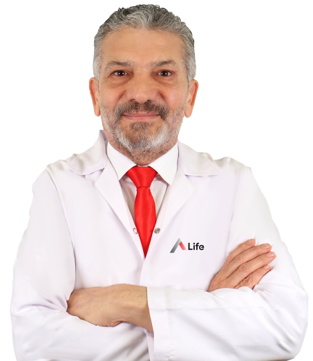
Op. Dr. Oğuz Fidan is a respected physician with extensive knowledge and many years of experience in the field of eye diseases. Aiming to provide his patients with the most current and effective treatment methods, Dr. Fidan has dedicated his professional life to serving humanity.
With a solid medical background, Dr. Fidan completed his medical education at Ankara University Faculty of Medicine, one of Turkey’s prestigious educational institutions.
He successfully completed his specialty training in eye diseases at Vehbi Koç Eye Specialty Hospital, a significant center in eye health, earning his expertise in ophthalmology.
Dr. Fidan has gained extensive experience by serving in various healthcare institutions throughout his professional career. He began his career as the Chief Physician at the SSK Ulucanlar Eye Bank, where he provided significant services for many years.
In addition to his medical education, Dr. Fidan's studies in law and philosophy of religion demonstrate that he is a multifaceted individual with a broad knowledge base and a wide worldview. This background contributes to offering a more comprehensive approach to his patients.
Currently continuing his work within A Life Sağlık Grubu, Dr. Fidan aims to provide high-quality healthcare services by utilizing his professional experience and knowledge. He is dedicated to resolving eye health issues and improving the quality of life for his patients.
As A Life Sağlık Grubu, we are pleased to offer you the expertise and experience of Op. Dr. Oğuz Fidan in eye health, especially in complex conditions such as optic atrophy.
Education Type | Institution
Medical Education | Ankara University Faculty of Medicine
Specialization | Vehbi Koç Eye Specialty Hospital
Institution
Ankara Occupational and Environmental Diseases Hospital
Ankara Etlik Specialty Training and Research Hospital
Ankara Çubuk Halıl Şivgin State Hospital
Private 34 Hospital
Private Veni Vidi Mamak Hospital
Private Natomed Hospital
Private Yüzüncüyıl Hospital
A Life Hospital Pursaklar
As an eye disease specialist, helping you with all issues related to your eye and vision health is my priority. My expertise includes diagnosing a wide range of diseases affecting the eye and vision system, planning treatments, and performing surgical interventions when necessary.
Some of the areas closely related to your eye health and my special interests are:
Refractive Errors
Myopia (nearsightedness): Inability to clearly focus on distant objects.
Hyperopia (farsightedness): Inability to clearly focus on close objects.
Astigmatism: Blurred vision at both near and far due to the eye focusing light unevenly on different axes.
Presbyopia: Age-related decline in near vision ability.
Cataract
Clouding of the eye lens causing decreased vision.
Glaucoma
A disease usually related to increased intraocular pressure that can cause optic nerve damage and vision loss.
Retinal Diseases
Macular Degeneration: A disease affecting the central part of the retina, common with aging.
Diabetic Retinopathy: Retinal damage caused by diabetes.
Retinal Detachment: Separation of the retina from the underlying tissue.
Retinitis Pigmentosa: A genetically inherited retinal disease.
Corneal Diseases
Keratoconus: Cone-shaped protrusion of the cornea.
Corneal Infections: Infections caused by bacteria, viruses, or fungi on the cornea.
Corneal Dystrophies: Hereditary corneal disorders.
Eyelid and Ocular Surface Diseases
Blepharitis: Inflammation of the eyelids.
Dry Eye: Insufficient tear production or poor tear quality.
Conjunctivitis: Inflammation of the conjunctiva, the transparent layer covering the eye.
Ptosis: Drooping of the eyelid.
Tear duct obstruction.
Neuro-ophthalmology
Diseases related to the optic nerve and visual pathways.
Strabismus and Pediatric Eye Diseases
Misalignment of the eyes (strabismus).
Amblyopia (lazy eye).
Other pediatric eye disorders.
Uveitis
Inflammation of one or more of the inner layers of the eye.
Eye Trauma
Eye injuries.
To protect and improve your eye health, I am pleased to offer you the most advanced eye surgical treatment options provided by modern medicine and technology. Surgical treatment of eye diseases covers a wide range from common conditions like cataracts, correction of refractive errors, to serious retinal problems. These treatments play a crucial role in restoring your vision and improving your quality of life.
With my expertise and experience, the main eye surgery areas I can offer you include:
Refractive Surgery (Glasses-Free Surgery)
LASIK (Laser-Assisted In Situ Keratomileusis): One of the most commonly used laser eye surgeries. A flap is created on the cornea and laser is applied.
PRK (Photorefractive Keratectomy): The surface epithelial layer of the cornea is removed and laser is applied.
LASEK (Laser Epithelial Keratomileusis): A combination of PRK and LASIK.
SMILE (Small Incision Lenticule Extraction): A minimally invasive method involving a small incision in the cornea to remove tissue.
Phakic Intraocular Lens Implantation: An additional artificial lens is implanted inside the eye without removing the natural lens.
These methods are used to correct myopia, hyperopia, and astigmatism.
Cataract Surgery
Phacoemulsification: A small incision is made, the cloudy lens is broken up with ultrasound waves and removed, and an artificial intraocular lens is implanted. This is the gold standard in cataract treatment.
Glaucoma Surgery
Various surgical procedures to reduce intraocular pressure and slow glaucoma progression, including trabeculectomy, tube implantation, and minimally invasive glaucoma surgery (MIGS).
Retina and Vitreous Surgery
Vitreoretinal Surgery: A set of surgical techniques used to treat retinal detachment, macular hole, vitreous hemorrhage, diabetic retinopathy, and other retina and vitreous diseases. These surgeries are performed under a microscope using specialized instruments.
Corneal Surgery
Corneal Transplantation (Keratoplasty): Replacing the diseased cornea with a healthy donor cornea. Different types exist (e.g., full-thickness corneal transplant, lamellar keratoplasty).
Corneal Cross-Linking: A method used to halt corneal shape disorders like keratoconus.
Oculoplastic Surgery
Surgery related to eyelids, tear ducts, orbit (eye socket), and surrounding facial tissues. Includes ptosis correction, entropion, ectropion, tear duct obstruction, eye tumors, and cosmetic eyelid surgeries (blepharoplasty).
Other Eye Surgery Procedures
Strabismus Surgery: Correction of misalignment caused by imbalance of eye muscles.
Pterygium Surgery: Removal of tissue growing from the white part of the eye onto the cornea.
Your eye health is very important to me. Therefore, I work meticulously to accurately diagnose any issues you experience with your eyes and to determine the most suitable treatment methods for you. The diagnosis process for eye diseases is not just a single test; it is a holistic approach formed by our communication with the patient, a comprehensive physical examination, and the evaluation of various advanced techniques together.
When necessary, based on this evaluation, we use various tests and imaging methods that provide more detailed information.
These tests are carefully selected and applied according to the specific needs of our patient.
Eyelid Examination
Conditions such as swelling, redness, drooping (ptosis), and abnormal positions (entropion, ectropion) of the eyelids are examined.
Eye Movement Examination
The function of the eye muscles and the coordination of the eyes are assessed.
Conjunctiva Examination
Redness, swelling, discharge, and other abnormalities of the conjunctiva, the membrane covering the white part of the eye (sclera) and the inner surfaces of the eyelids, are evaluated.
Cornea Examination
The transparency, shape, and surface smoothness of the cornea are assessed.
Pupil Examination
The size, shape, reaction to light, and symmetry of the pupils are examined.
Anterior Chamber Examination
The depth and clarity of the fluid-filled space in the front part of the eye (anterior chamber) are checked.
Iris and Lens Examination
The color and shape of the iris and the clarity of the lens are evaluated.
Visual Acuity Test
One of the most commonly used eye tests. The patient is asked to read specific letters, numbers, or symbols from different distances. Visual clarity is determined.
Refraction (Glasses) Examination
Determines the eyeglass prescription that provides the best visual acuity for the patient. Refraction can be performed using specialized devices (phoropter, retinoscope) or computer-assisted systems (autorefractor). Refractive errors such as myopia (nearsightedness), hyperopia (farsightedness), and astigmatism are detected.
Intraocular Pressure Measurement (Tonometry)
Measures the pressure inside the eye. Important for diagnosing and monitoring glaucoma (eye pressure disease).
Biomicroscopic Examination
Using a high-magnification special microscope (biomicroscope, slit lamp), the anterior and posterior segment structures of the eye are examined in detail. Eyelids, cornea, iris, lens, vitreous, and retina are inspected.
Fundus Examination (Ophthalmoscopy)
The back of the eye (retina, optic nerve) is examined by dilating the pupil (using eye drops or special devices). The retina, macula (yellow spot), blood vessels, and optic nerve are evaluated.
Additional Diagnostic Methods (When Needed)
Fluorescein Angiography (FFA) or Indocyanine Green Angiography (ICGA): Tests used to examine the blood vessels of the retina or choroid (layer under the retina).
Optical Coherence Tomography (OCT): Used to obtain detailed cross-sectional images of the retina, macula, and optic nerve.
Visual Field Test: Conducted to determine the limits of vision and detect any visual field loss.
Corneal Topography: Maps the surface curvature and thickness of the cornea.
Other Tests: Color vision tests, contrast sensitivity tests, strabismus tests, electrophysiological tests, etc.
Elvan Mahallesi 1934/4 Etimesgut/Ankara
+90 (850) 888 54 33
Aydınlıkevler, Uzayan Sk. No:99, 06130 Altındağ/Ankara
0850 850 54 33
Fatih Mah, Yavuz Bulv, No:15 Pursaklar / Ankara
+90 (850) 888 54 33
7/24 tüm soru ve sorunlarınız için buradayız.
Lütfen size ulaşabilmek için aşağıdaki alanları doldurunuz
Başlangıç Tarihi:
Bitiş Tarihi: